
🫀 What is PDA?
Patent Ductus Arteriosus is a persistent opening between the aorta and pulmonary artery that should normally close shortly after birth. When it remains open, oxygen-rich blood flows abnormally into the lungs, causing overload.

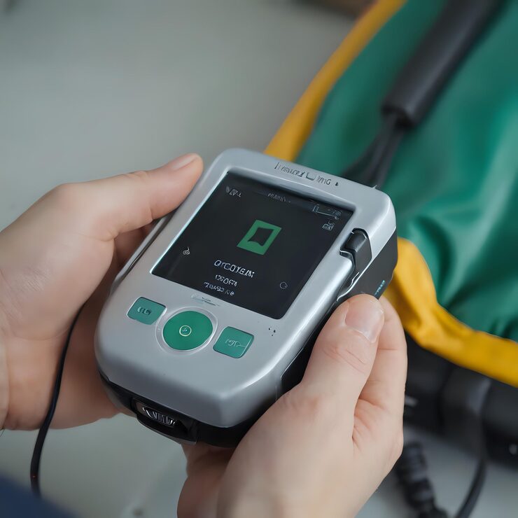
PDA device closure is a safe, catheter-based procedure that seals the abnormal connection without open-heart surgery and allows rapid recovery.
Types of PDA
- Small / Silent PDA
- Moderate PDA
- Large PDA
- Tubular, Elongated PDAs
- Krichenko Types A–E (angiographic classification)
Who Needs PDA Closure?
- Babies with failure to thrive
- Frequent pneumonia or respiratory issues
- Continuous murmur on examination
- Left heart enlargement on Echo
- Large PDA causing increased lung blood flow
- Endocarditis prevention in moderate/large PDA
Procedure Overview: Transcatheter PDA Device Closure
Step-by-step:
- Local or general anesthesia (depending on age)
- A small catheter is inserted through femoral vein/artery
- PDA is crossed using a guidewire
- A coil or occluder device is deployed
- Device seals PDA completely
- Catheters removed — no stitches
- ⏱ Duration: 30–60 minutes
- 🛏 Hospital Stay: Same day or 1 day
Benefits
- No open surgery
- Quick recovery
- Very high success rate (>99%)
- No scar
- Normal activity within days
Risks (Very Rare)
- Device embolization (<1%)
- Mild residual leak (usually closes within weeks)
- Vessel injury or bleeding
- Allergic reaction to contrast dye
PDA Closure in Infants & Children
- Safe even in small infants
- Coil devices for tiny PDAs
- Amplatzer duct occluder (ADO) for large PDAs
- Rapid improvement in breathing and weight gain
PDA Closure in Adults
Required when:
- Symptom causing PDA
- Enlarged left chambers
- Risk of endarteritis
VSD Device Closure (Ventricular Septal Defect)
🫀 What is VSD?
A Ventricular Septal Defect (VSD) is a hole in the wall separating the two lower chambers of the heart (ventricles). It causes oxygen-rich blood to mix with oxygen-poor blood.
Types of VSD
- Perimembranous VSD (most common)
- Muscular VSD (best suited for device closure)
- Subpulmonic / Outlet VSD
- Inlet VSD
Who Needs VSD Device Closure?
- Failure to thrive in infants
- Frequent respiratory infections
- Large left-to-right shunt
- Left heart chamber enlargement
- Pulmonary hypertension (early, reversible)
- Previous infective endocarditis
Procedure Overview: Transcatheter VSD Device Closure
Step-by-step:
- General anesthesia (especially in children)
- Catheter inserted through the femoral vein/artery
- Crossing the VSD using guidewires
- A double-disc nitinol occluder device is deployed
- Device seals the VSD completely
- No stitches — only a small puncture site
- ⏱ Duration: 60–120 minutes
- 🛏 Hospital Stay: 1–2 days